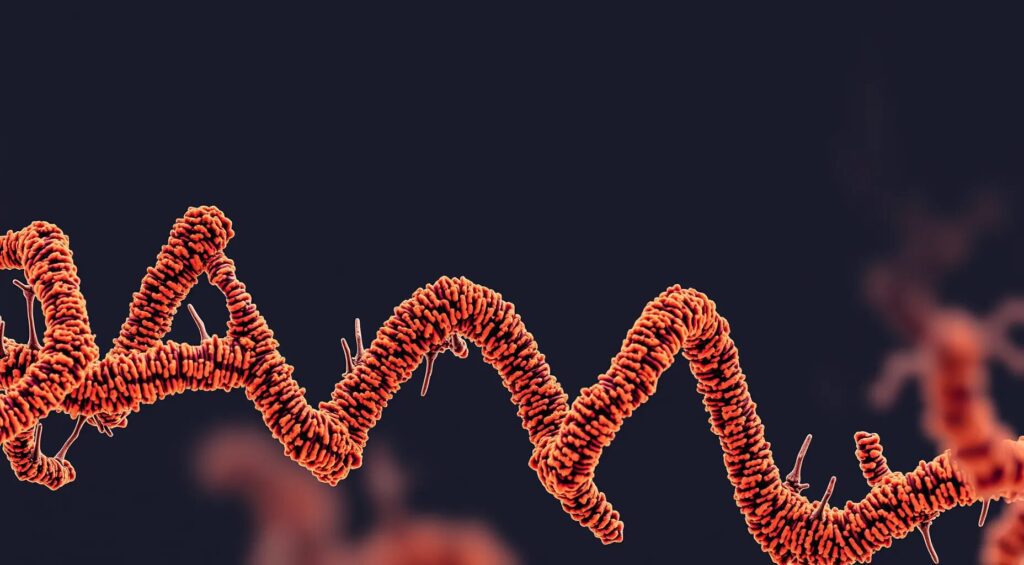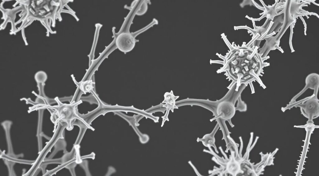
Electron microscopy, since its inception, has been a cornerstone in the exploration of biological structures at a nanoscale level. Traditional electron microscopes rely on a beam of electrons instead of light to illuminate samples, allowing scientists to visualize structures at resolutions down to the atomic level. However, despite these incredible capabilities, early electron microscopy was limited by several factors, including sample preparation challenges, the time-consuming nature of data collection, and the immense complexity of image interpretation.
These limitations began to dissolve with the advent of AI, particularly machine learning and deep learning algorithms, which have enabled a leap from merely capturing images to extracting meaningful biological information. AI doesn’t just improve image clarity; it fundamentally changes the way we interpret those images, transforming them into actionable biological insights.
AI in Structural Biology: Breaking Down Complex Systems
In structural biology, the study of macromolecules like proteins, nucleic acids, and their complexes is critical to understanding the biochemical processes that sustain life. Before the integration of AI, resolving the 3D structures of these molecules was a painstaking process, often requiring years of labor-intensive work.
Cryo-Electron Microscopy (Cryo-EM) has emerged as a transformative technology in this field, allowing scientists to capture images of frozen-hydrated specimens at near-atomic resolution. However, the real breakthrough came when AI was introduced to analyze the vast amounts of data generated by cryo-EM. AI-driven algorithms can now automatically sort through millions of particle images, classifying them into groups based on similarity, and reconstructing high-resolution 3D structures with unprecedented speed and accuracy.
One notable example is the AI-driven reconstruction of the ribosome, the molecular machine responsible for protein synthesis in cells. Prior to AI, understanding the ribosome’s intricate structure and function was a formidable challenge. With AI, scientists have mapped the ribosome at atomic resolution, revealing its dynamic conformational states during protein synthesis. This has not only deepened our understanding of fundamental biological processes but has also identified potential targets for antibiotics, offering new avenues for drug development.
AI and Protein Folding: Predicting the Future of Medicine
Another groundbreaking application of AI in biological imaging is in the realm of protein folding. The three-dimensional shape of a protein dictates its function, and misfolding can lead to diseases such as Alzheimer’s, Parkinson’s, and various forms of cancer. Traditionally, determining a protein’s structure required experimental methods like X-ray crystallography or NMR spectroscopy, which could take years and might not always be successful.
Enter AlphaFold, an AI system developed by DeepMind, which has revolutionized the field by predicting protein structures with atomic accuracy based solely on their amino acid sequences. While AlphaFold’s primary contribution is computational, its predictions can be validated and further refined using AI-enhanced cryo-EM. This synergy allows researchers to not only predict but also visualize protein structures and their dynamic interactions within the cell.
For instance, in the case of SARS-CoV-2, the virus responsible for COVID-19, AI predicted the structure of various viral proteins, which was crucial in understanding how the virus invades human cells. These predictions, validated by cryo-EM, played a critical role in designing vaccines and therapeutic antibodies at record speed.

AI in Cellular Imaging: Unveiling the Inner Workings of Cells
Beyond individual proteins, AI-enhanced electron microscopy is also transforming our understanding of whole cellular systems. 3D Cellular Tomography is a technique that allows scientists to capture a series of 2D electron microscope images from different angles and then reconstruct them into a 3D model of the cell. However, interpreting these complex datasets manually is a Herculean task.
AI comes to the rescue by automating the segmentation and analysis of cellular components. Using deep learning, AI algorithms can identify and label different organelles, membranes, and other cellular structures with remarkable precision. This has led to new insights into how cells function at a systems level.
A fascinating case is the use of AI to study the synapses in the brain. Synapses are the junctions between neurons where communication occurs, and understanding their structure is key to unraveling the mysteries of the brain. AI-enhanced electron microscopy has allowed scientists to map entire neural circuits at nanometer resolution, revealing the detailed architecture of synapses and how they change in diseases like Alzheimer’s and schizophrenia.
AI and Viral Imaging: A New Weapon in the Fight Against Pandemics
The fight against infectious diseases has always been a race against time. AI-enhanced electron microscopy has proven to be a critical weapon in this battle, particularly in the study of viruses. Viruses are small, rapidly mutating, and often structurally complex, making them difficult targets for traditional imaging techniques.
AI has changed the game by enabling the rapid analysis and interpretation of viral structures. During the COVID-19 pandemic, researchers used AI-driven cryo-EM to rapidly solve the structure of the SARS-CoV-2 spike protein, the key molecule the virus uses to enter human cells. This structural information was shared globally and became the foundation for the design of vaccines and antiviral drugs.
But the story doesn’t end with SARS-CoV-2. AI-enhanced imaging is now being used to study a wide range of viruses, including influenza, HIV, and Ebola. These insights are helping to identify vulnerabilities in viral structures that can be exploited for therapeutic intervention, offering hope in the fight against both current and future pandemics.
Beyond Imaging: AI as a Discovery Tool
While the primary role of AI in electron microscopy has been to enhance imaging, its potential goes far beyond visualization. AI is increasingly being used as a discovery tool, helping to generate hypotheses and guide experiments in ways that were previously unimaginable.
For example, AI can analyze vast datasets generated by electron microscopy to identify patterns that humans might miss. In studying complex biological systems, AI algorithms can suggest new relationships between structures and functions, driving the discovery of previously unknown biological mechanisms.
A recent example comes from cancer research, where AI was used to analyze electron microscopy images of tumor cells. The AI identified previously unrecognized substructures within the cells that are associated with aggressive tumor behavior. These findings have opened up new avenues for understanding how cancer spreads and for developing targeted therapies.
Scientists have developed an AI-driven method that enhances the capabilities of electron microscopy. By integrating machine learning algorithms with traditional microscopy techniques, this new approach refines image quality and boosts resolution. It’s akin to putting on a pair of glasses after years of blurred vision—suddenly, everything is sharper, clearer, and more detailed.
— Their findings have recently been published in Nature Communications
Challenges and Future Directions
While the potential of AI-enhanced electron microscopy is vast, there are still challenges to overcome. One of the main issues is the need for large, high-quality datasets to train AI algorithms. In many areas of biology, such datasets are scarce or difficult to obtain. Moreover, as AI models become more complex, the interpretability of their results can diminish, leading to a “black box” problem where it’s unclear how the AI arrived at its conclusions.
However, the field is rapidly advancing, and efforts are being made to address these challenges. Open-source initiatives and collaborations between research institutions are helping to build the large, diverse datasets needed for training AI models. At the same time, researchers are developing new AI techniques that are more interpretable and better suited to the unique challenges of biological imaging.
Looking ahead, we can expect AI to play an even more integral role in biological discovery. As AI algorithms become more sophisticated and integrated with other technologies, such as quantum computing and advanced molecular simulation, the possibilities for biological imaging will continue to expand. This could lead to breakthroughs in our understanding of life at the molecular level, new treatments for diseases, and perhaps even the ability to manipulate biological systems with unprecedented precision.
Conclusion: The New Frontier of Biological Imaging
AI-enhanced electron microscopy represents a new frontier in biological imaging, one where the boundaries between seeing and understanding are increasingly blurred. As AI continues to evolve, it is transforming how we study life, enabling us to explore biological systems in unprecedented detail.
From the intricate folds of proteins to the complex interactions within cells, and from the deadly architecture of viruses to the neural circuits of the brain, AI is opening up new worlds of discovery. The fusion of AI and electron microscopy is more than just a technological advancement—it’s a paradigm shift that is reshaping our understanding of biology.
In this era of rapid scientific progress, the journey from pixels to proteins is not just a metaphor for technological advancement; it is a reality that promises to unlock the deepest secrets of life itself. And as we continue to push the boundaries of what is possible with AI, there is no telling what new insights and discoveries await us on the horizon.
Peer-reviewed journals and other resources that delve into the topics discussed:
- DeepMind’s AlphaFold and Protein Folding
Reference: Jumper, J., Evans, R., Pritzel, A., et al. (2021). Highly accurate protein structure prediction with AlphaFold. Nature, 596, 583–589.
DOI: 10.1038/s41586-021-03819-2 - Cryo-Electron Microscopy and Structural Biology
Reference: Cheng, Y. (2018). Single-particle cryo-EM—How did it get here and where will it go. Science, 361(6405), 876-880.
DOI: 10.1126/science.aat4346 - AI in Cellular Imaging
Reference: Ebrahim, M., Tassone, G., & Wright, G. (2020). The impact of AI on electron microscopy: Understanding macromolecular structures from cells to viruses. Journal of Structural Biology, 211(1), 107540.
DOI: 10.1016/j.jsb.2020.107540 - AI and Viral Structure Analysis
Reference: Walls, A. C., Park, Y. J., Tortorici, M. A., Wall, A., McGuire, A. T., & Veesler, D. (2020). Structure, Function, and Antigenicity of the SARS-CoV-2 Spike Glycoprotein. Cell, 181(2), 281-292.e6.
DOI: 10.1016/j.cell.2020.02.058 - AI and Cancer Research Using Electron Microscopy
Reference: Liu, Y., Gadepalli, K., Norouzi, M., et al. (2017). Detecting Cancer Metastases on Gigapixel Pathology Images. Journal of the American Medical Association, 318(22), 2199-2210.
DOI: 10.1001/jama.2017.14585

